Name:____________________________________________________________ Per:_______ Date:_______________
A2 The Human Brain Study Guide
Introduction:
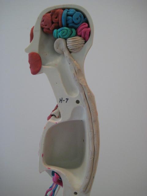
The brain allows us to move, to breathe, to make decisions, to solve problems, to feel emotions and to interact with the world around us. This 3lb, jelly like organ is often compared to a computer, but the brain is so much more complicated than even the biggest, fastest supercomputer- thanks to the 100 billion nerve cells that keep our body functioning (that’s more nerve cells than stars in the galaxy!). The brain is the key to communication in the human body. This organ not only allows your systems to communicate with one another, but also allows you to communicate with and respond to your surroundings.
A2 - Build a Brain & Neurology Scenarios
Directions:
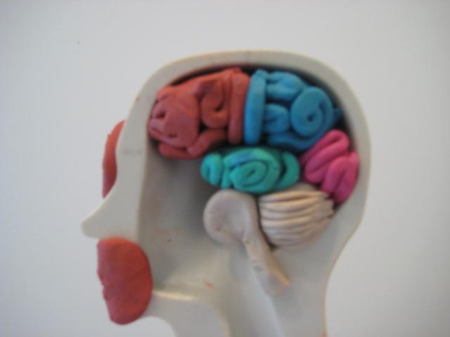
In this activity, you will design and then build a model brain. As you build, you will begin to see connections between specific parts of the brain and specific actions, processes and functions that make us human. Use the resource packet along with the link located on the blog to design a brain. You will need to demonstrate that you know regions of the brain within the key terms and solve two scenarios.

1.Design Brain Directions: Click on the links (feel free to do your own research) to get a better sense of the brain.
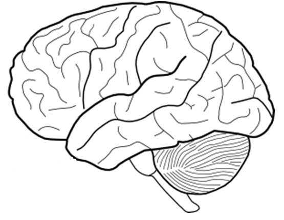
2. Draw and color code the following brain regions: Frontal lobe, Parietal, Temporal, Parietal, Occipital Cerebellum, and brainstem. Go to the neuro-skills website in order to create a color code key for different part of the lobes of the brain.
3. Directions to build model brain: Use the links above and the resource packet directions to build your very own brain using the key terms #1 first. Pay attention to detail and locations since you will need to demonstrate you know their anatomical location and fill in the graphic organizer to earn a stamp.


Make sure you use anatomical terminology (do some research) to fill in the graphic organizer.
Region of the Brain | Location (Use anatomical terminology) | Primary Function |
Cerebrum | ||
Frontal Lobe | ||
Parietal lobe | ||
Occipital lobe | ||
Temporal Lobe | ||
Cerebellum | ||
Brain Stem |
4. Neuro-Scenarios Directions: Answer the questions below in full sentences. These scenarios are intended for you to research and decide the best answer (using the resources above). Remember there will be more than one correct answer and you must provide an explanation with each answer.
- A man slips, falls and bangs the back of his head on the tile floor. The doctor tells him he has some minor swelling in the occipital lobe of his cerebrum. His doctor sends him home to rest and tells him that if there are any changes, he should come back in. What changes would be worrisome, given the area that was injured?

- A car going at 25mph along NE Broadway and Sandy hits a bicyclist. The person was not wearing a helmet and is rushed into the OHSU emergency room. He complains that he has trouble walking and trembles and shakes trying to hold anything. He also complains that he has lost sensation in right leg although he was able to move it. What are the possible injuries occurring to the brain?

- A patient at Legacy has seizures that occur along one side of his brain and travels to the other brain. You the doctor have to make some decisions in how to treat his seizures by either surgery or medication. What would you do and explain your rationale below (feel free to do research on this).

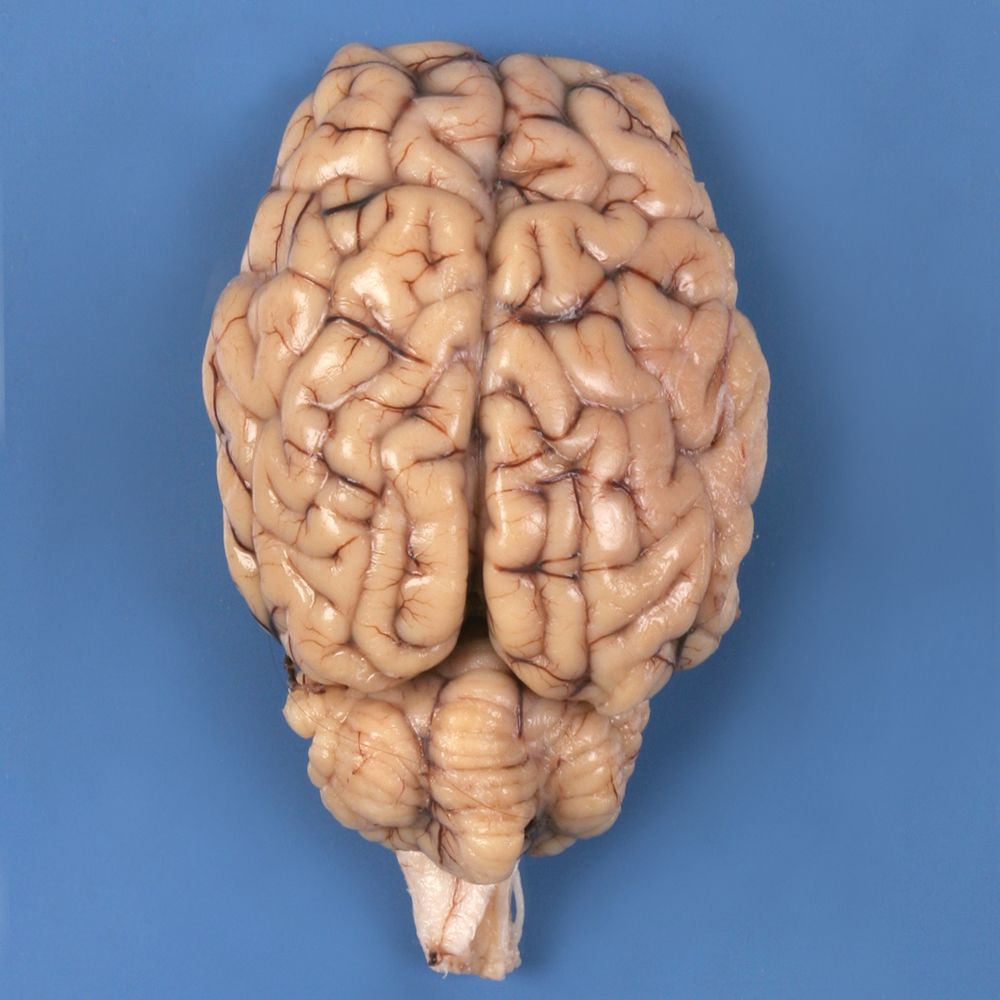
Activity 2: Sheep Brain Dissection Lab
Directions: You will be using a dissecting tray, along with sharp objects. You will need to use gloves and have goggles on. Remember these brains are fixed with formaldehyde and are toxic/life threatening. Please use caution when dissecting.
1. Obtain a dissection pan, a set of dissecting tools, some toothpicks and labels for tape flags, as well as a sheep brain. Assemble 20 tape flags in your groups using the toothpicks and adhesive labels. Assemble more as needed as you complete the initial dissection as well as your map.
Cerebrum Directions: First look at the brain and use Key Terms #1 to put toothpick where you think each anatomical structure is located. I will come around and ask three random anatomical structures and their function.
Anatomical Structures | Toothpick # | Function |
Frontal lobe | ||
Parietal lobe | ||
Occipital lobe | ||
Temporal lobe | ||
You Pick: __________________ | ||
You Pick: __________________ | ||
You Pick: __________________ | ||
Medulla |
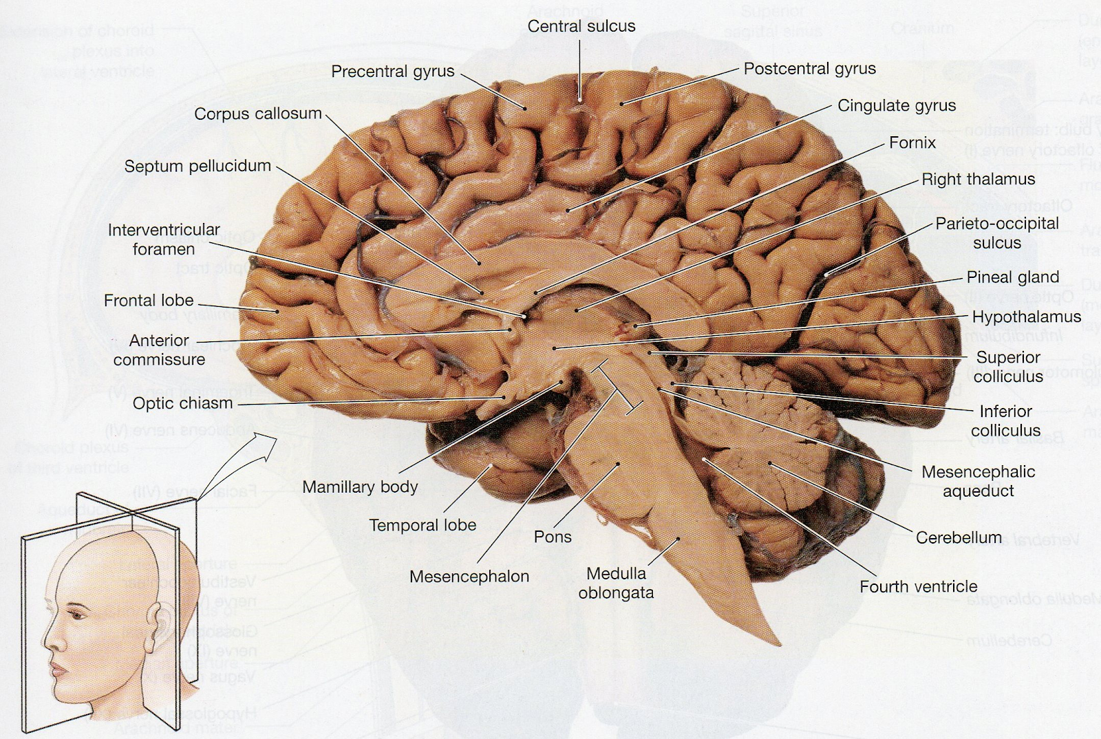
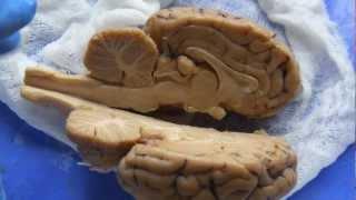
Sagittal Directions: Create a midsagittal cut. Hold the brain level flat and cut along the longitudinal fissure. Use a toothpick to label the anatomical areas. Fill out the graphic organizer below.
Anatomical Structures | Toothpick # | Function |
Corpus Callosum | ||
Thalamus | ||
Hypothalamus | ||
Pons | ||
Cerebellum
| ||
Lateral Ventrical |
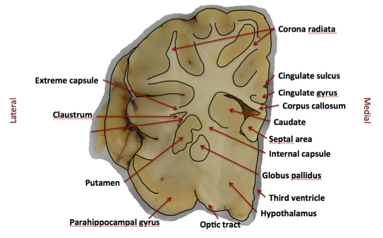
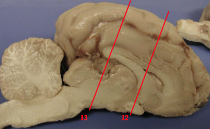
Coronal Directions: Create a coronal cut. Hold the brain level flat and cut along the brain as illustrated below. Use the toothpick to label the particular areas below, and use the image on the next page to help you identify particular regions in your brain. Fill out the graphic organizer below (research).
Anatomical Structures | Toothpick # | Function |
Optic Chiasma | ||
Internal Capsule | ||
Caudate Nucleus | ||
Globus Pallidus | ||
You Pick:
| ||
You Pick:
|