7.9 The Urinary System
Learning Objectives
- Describe the parts of urinary system.
- Outline how the kidneys filter blood.
- Describe what urine is and how it is formed.
Introduction
One of the most important ways your body maintains homeostasis is by keeping the right amount of water and salts inside your body. If you have too much water in your body, your cells can swell and burst. If you have too little water in your body, your cells can shrivel up like an old apple. Both of these situations occur because of the process of osmosis! Either extreme can cause illness and death of cells, tissues, and organs. The organs of your urinary system help to keep the correct balance of water and salts within your body.
Guided Learning
The Urinary System
The urinary system is the organ system that makes, stores, and gets rid of urine. It includes:
- Two kidneys
- Two ureters
- One bladder
- One urethra
The urinary system is shown in Figure below.
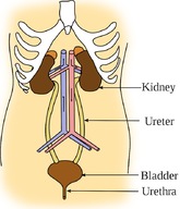
The kidneys filter the blood that passes through them and the urinary bladder stores the urine until it is released from the body.
Organs of the Urinary System
- As you can see from Figure above, the kidneys are two bean-shaped organs. Kidneys filter and clean the blood and form urine. They are about the size of your fists and are found near the middle of the back, just below your rib cage.
- Ureters are tube-shaped and bring urine from the kidneys to the urinary bladder.
- The urinary bladder is a hollow and muscular organ. It is shaped a little like a balloon. It is the organ that collects urine.
- Urine leaves the body through the urethra.
What is Urine?
Urine is a liquid that is formed by the kidneys when they filter wastes from the blood. Urine contains mostly water, but also contains salts and nitrogen-containing molecules. The amount of urine released from the body depends on many things. Some of these include the amounts of fluid and food a person consumes and how much fluid they have lost from sweating and breathing. Urine ranges from colorless to dark yellow, but is usually a pale yellow color. Light yellow urine contains mostly water. The darker the urine, the less water it contains.
The urinary system also removes a type of waste called urea from your blood. Urea is a nitrogen-containing molecule that is made when foods containing protein, such as meat, poultry, and certain vegetables, are broken down in the body. Urea and other wastes are carried in the bloodstream to the kidneys, where they are removed and form urine.
How the Kidneys Filter Wastes
The kidneys are important organs in maintaining homeostasis. Kidneys perform a number of homeostatic functions.
- They maintain the volume of body fluids.
- They maintain the balance of salt ions in body fluids.
- They excrete harmful nitrogen-containing molecules, such as urea, ammonia, and uric acid.
There are many blood vessels in the kidneys, as you can see in Figure below. The kidneys remove urea from the blood through tiny filtering units called nephrons. Nephrons are tiny, tube-shaped structures found inside each kidney. A nephron is shown in Figure below. Each kidney has up to a million nephrons. Each nephron collects a small amount of fluid and waste from a small group of capillaries.
If the body is in need of more water, water is removed from the fluid inside the nephron and is returned to the blood. The fluid within nephrons is carried out into a larger tube in the kidney called a ureter, which you can see in Figure below. Urea, together with water and other wastes, forms the urine as it passes through the nephrons and the kidney.

Structures of the kidney: fluid leaks from the capillaries and into the nephrons where the fluid forms urine then moves to the ureter and on to the bladder.
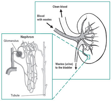
The location of nephrons in the kidney. The fluid collects in the nephron tubules, and moves to the bladder through the ureter.
Formation of Urine
The process of urine formation is as follows:
- Blood flows into the kidney through the renal artery, shown in Figure below. The renal artery connects to capillaries inside the kidney. Capillaries and nephrons lie very close to each other in the kidney.
- The blood pressure within the capillaries causes water, salts, sugars, and urea to leave the capillaries and move into the nephron.
- The water and salts move along through the tube-shaped nephron to a lower part of the nephron.
- The fluid that remains in the nephron at this point is called urine.
- The blood that leaves the kidney in the renal vein has much less waste than the blood that entered the kidney.
- The urine is collected in the ureters and is moved to the urinary bladder, where it is stored.
Nephrons filter about ¼ cup of body fluid per minute. In a 24-hour period, nephrons filter 180 liters of fluid, and 1.5 liters of the fluid is released as urine. Urine enters the bladder through the ureters. Similar to a balloon, the walls of the bladder are stretchy. The stretchy walls allow the bladder to hold a large amount of urine. The bladder can hold about 1½ to 2½ cups of urine, but may also hold more if the urine cannot be released immediately.
How do you know when you have to urinate? Urination is the process of releasing urine from the body. Urine leaves the body through the urethra. Nerves in the bladder tell you when it is time to urinate. As the bladder first fills with urine, you may notice a feeling that you need to urinate. The urge to urinate becomes stronger as the bladder continues to fill up.
Brain Control
The kidneys never stop filtering waste products from the blood, so they are always producing urine. The amount of urine your kidneys produce is dependent on the amount of fluid in your body. Your body loses water through sweating, breathing, and urination. The water and other fluids you drink every day help to replace the lost water. This water ends up circulating in the blood because blood plasma is mostly water.
The filtering action of the kidneys is controlled by the pituitary gland. The pituitary gland is about the size of a pea and is found below the brain, as shown in Figure below. The pituitary gland is also part of the endocrine system. The pituitary gland releases hormones, which help the kidneys to filter water from the blood.
The movement of water back into blood is controlled by a hormone called antidiuretic hormone (ADH). ADH is released from the pituitary gland in the brain. One of the most important roles of ADH is to control the body’s ability to hold onto water. If a person does not drink enough water, ADH is released. It causes the blood to reabsorb water from the kidneys. If the kidneys remove less water from the blood, what will the urine look like? It will look darker, because there is less water in it.
When a person drinks a lot of water, then there will be a lot of water in the blood. The pituitary gland will then release a lower amount of ADH into the blood. This means less water will be reabsorbed by the blood. The kidneys then produce a large volume of urine. What color will this urine be?
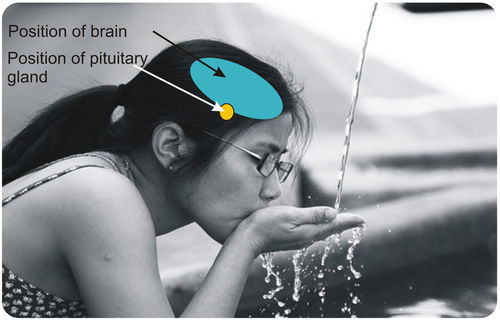
The pituitary gland is found directly below the brain and releases hormones that control how urine is produced.
Summary
- The organs of the urinary system remove wastes. They also maintain the proper levels of water, salts, and nutrients in the body.
- The urinary system is made up of the kidneys, the ureters, the bladder, and the urethra.
- The filtering parts of the kidneys are the nephrons.
- Water and waste molecules move out of the blood capillaries and into the nephrons. Most of the water returns to the blood.
- Urine collects in the nephron and moves to the urinary bladder through the ureters.
- The filtering action of the kidneys is controlled by the pituitary gland.
- ADH is the hormone released by the pituitary gland and controls the how water is reabsorbed by the blood from the kidneys.
Vocabulary
kidney
An organ that filters and cleans the blood and forms urine; about the size of a fist and are found near the middle of the back, just below your rib cage.
nephron
A tiny, tube-shaped structure found inside each kidney that collects a small amount of fluid and waste from a small group of capillaries; up to a million nephrons are found in each kidney.
urinary bladder
A hollow and muscular organ that collects urine and is shaped like a little balloon.
urinary system
The organ system that makes, stores, and gets rid of urine; consists of kidneys, ureters, bladder, and urethra.
urination
The process of releasing urine from the body; urine leaves the body through the urethra and nerves in the bladder tell you when it is time to urinate.
urine
A liquid that is formed by the kidneys when they filter wastes from the blood; contains mostly water, but also contains salts and nitrogen-containing molecules.

Licensed under • Terms of Use • Attribution With additions made by the MN Partnership for Collaborative Curriculum.
")
[1] Urinary System by SEER / CK-12 / CC-BY-SA 3.0.
[2] Kidney Structures by PIOTR MICHAL JAWORSKI / CK-12 / CC-BY-SA 3.0.
[3] Nephron Ureter by NIH / CK-12 / CC-BY-SA 3.0.
[4] Pituitary by J.C. ROJAS / CK-12 / CC-BY-SA 3.0.