Malignant melanoma, recognized as the most severe form of skin cancer essentially develops as tumors arising from melanocytes (skin cells) that produce melanin - the pigment that provides skin with its color. Despite the infrequency of melanoma cases, the American Cancer Society cites that this type of skin cancer causes 75% of skin cancer deaths, particularly due to the nature of this cancer to metastasize to other regions of the body The American Cancer Society predicts that in the United States in 2013 alone, about 76,690 new cases will be diagnosed and about 9,480 people are expected to die of melanoma. On the global perspective, the World Health Organization cites that about 132,000 cases of melanoma develop annually. More pertinently, unlike many other cancers, melanoma can occur in both younger and older age groups, and its rates have been significantly rising in the past three decades.

Figure 1: Melanoma Mortality Rates in U.S. (2000-2004)
Source: National Cancer Institute
Figure 1: (Link: http://www.uspharmacist.com/content/d/health%20systems/c/33449/)
Essentially, while melanoma incidence rates continue to rise, the importance of early detection cannot be undermined. Generally, five year survival rates for patients with Stage I melanoma typically exceed 90 to 95%. However, as melanoma progresses as in later stages, these five year survival rates drop to less than 50%. Taking this into consideration, the earlier melanoma is diagnosed in its localized form (which typically in the epidermis), the greater the chance of survival becomes with a better prognosis.


Figure 2 | Figure 3 |
Figure 2 & 3: Early Stage Melanoma
Source: National Cancer Institute
Figure 2 Link: http://www.cancer.gov/cancertopics/pdq/treatment/melanoma/HealthProfessional/page1
Generally, the common issue with the detection of melanoma lies in the lack of time and resources in discerning if a dermatologist check-up is necessary in the event upon which a suspicious mole or mark appears on the skin. To target this issue, MCIT Indianapolis seeks to develop a Saccharomyces cerevisiae (yeast) biosensor that could be incorporated in a topical cream that has the ability to fluoresce in the presence of a potentially malignant melanoma tumor. The biosensor would be comprised of a yeast chassis containing an Fibroblast Growth Factor Receptor-1 (FGFR-1) expressed receptor and a plasmid consisting of the Renilla and Firefly Luciferase dual biobrick, that would serve as key components upon the detection and signaling of the presence of the a malignant melanoma tumor. As a biosensor, the device to be constructed will serve to carry out three general processes: detection of input, activation of signaling pathway, and activating response (output).
The first phase of the biosensor system is detection of input. In regards to the project, the input to be detected consists of mutated Basic Fibroblast Growth Factor (FGF2) cell surface proteins commonly located upon the surface of malignant melanoma tumors. Typically, in melanoma, FGF2 (BFGF) cell surface proteins are highly expressed along with their respective transmembrane receptors, the Fibroblast Growth Factor Receptor-1 molecules, or FGFR-1. In fact, one of the main characteristics of melanoma is a change from paracrine growth factor stimulation by surrounding keratinocytes in the skin to autocrine stimulation via dysregulated expression of growth factors and receptors on melanocytes. Previous studies have indicated that the proliferation, cell division, and cell survival depend upon FGF2 molecules, as well as FGF2 and FGFR-1 signaling, in which case this pathway has been known to set in motion a cascade of downstream signals through protein kinase pathways, as the MAPK pathway, so as to ultimately influence mitogenesis and differentiation. In the case of this project, an FGFR-1 biobrick will be used to express the FGFR-1 molecule on the yeast chassis, which will have the ability to detect FGF2 proteins on the surface of malignant melanoma tumors.
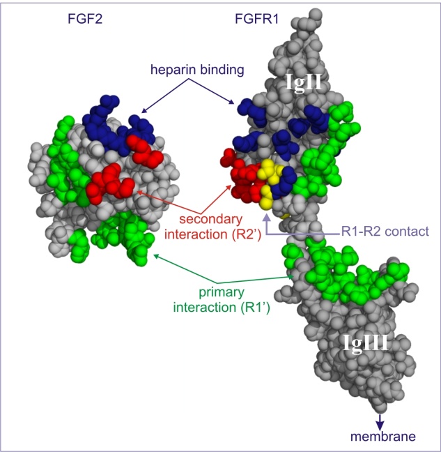
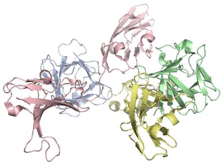
Figure 4 | Figure 5 |
Figure 4: Three-dimensional model of FGF2 (BFGF) protein Source: Diabetic retinopathy database | Figure 5: Binding of FGF2 and FGFR-1 Source: Institut Europée de Chimie et Biologie |

Figure 6 |
Figure 6: Three-dimensional model of FGF2 (BFGF) & FGFR-1 Complex Source: University of Cambridge, Department of Biochemistry |
Image Links:
Figure 4: http://diaretinopathydatabase.com/FGF2.html
Figure 5: http://www.cellbiol.net/ste/rpimages.php
Figure 6: http://www.sciencedirect.com/science/article/pii/S0959440X0000258X
The second phase of the biosensor system is activation of the signaling pathway. The signaling pathway is a cascade of events that is activated after over expressed FGF2 proteins from malignant melanocytes bind to the FGFR-1 receptor located on the yeast chassis. This cascade of events serves to induce expression of the Renilla and Firefly Luciferase dual biobrick that will eventually produce the fluorescent glow or the output in this case. The project itself will only address the detection of input and activating response phases, and not the activation of the signaling pathway phase, as this is a potential future endeavor to be researched.
The third phase of the biosensor system is the activating response (output), which represents the activation of the Firefly Luciferase and Renilla dual biobrick, which will encode for a reporter gene complex that qualitatively indicates the amount of FGF2 proteins present upon the surface of a potentially malignant melanoma tumor. Essentially, Luciferase represents a class of oxidative enzymes used in bioluminescence. In particular the firefly luciferase reaction produces a range of light that can vary between yellow-green light (550 nm) to red light (620 nm). In the biosensor system, once FGF2 molecules on the surface of the malignant melanoma detectors have bound to the FGFR-1 receptors of the biosensor, a resulting signaling pathway will induce arabinose expression which will then induce the Firefly Luciferase and Renilla biobrick to emit a glow based upon how many FGF2 molecules were detected. As more FGF2 molecules are detected, the glow emitted will increase in its intensity, signifying the potential severity of the potential malignant melanoma tumor.
Figure 7 |
Figure 7: Three-dimensional model of Firefly Luciferase Source: Blackett Laboratory, Imperial College, UK |
