Cells
Lesson 3.5
Cell division in eukaryotic cells and cancer
3.5.1 Introduction to cell division
It is obvious that you are not the same size as the day of your birth. When you break a bone, or cut yourself, the bone and the skin heal. Sometimes when you scratch your skin, white flakes of dead skin fall off. The question is why?
Cells are restricted to a particular size due to the cell membrane’s surface area to cell volume ratio. This ratio needs to be maintained in order for cells to function effectively. Typically, the volume (what’s inside the cell) will increase more quickly than the cell membrane’s surface area. That situation eventually creates a problem for the cell to efficiently obtain nutrients and remove wastes. Division of the cell allows for a shift back to an acceptable surface area to volume ratio. How frequently a particular cell divides varies with its type and function within the organism. Some cells, such as certain nerve cells in humans, do not reproduce.
The answer to this dilemma lies with the process of the mitosis, which is part of the larger cell cycle as seen in Figure 3.5.1.
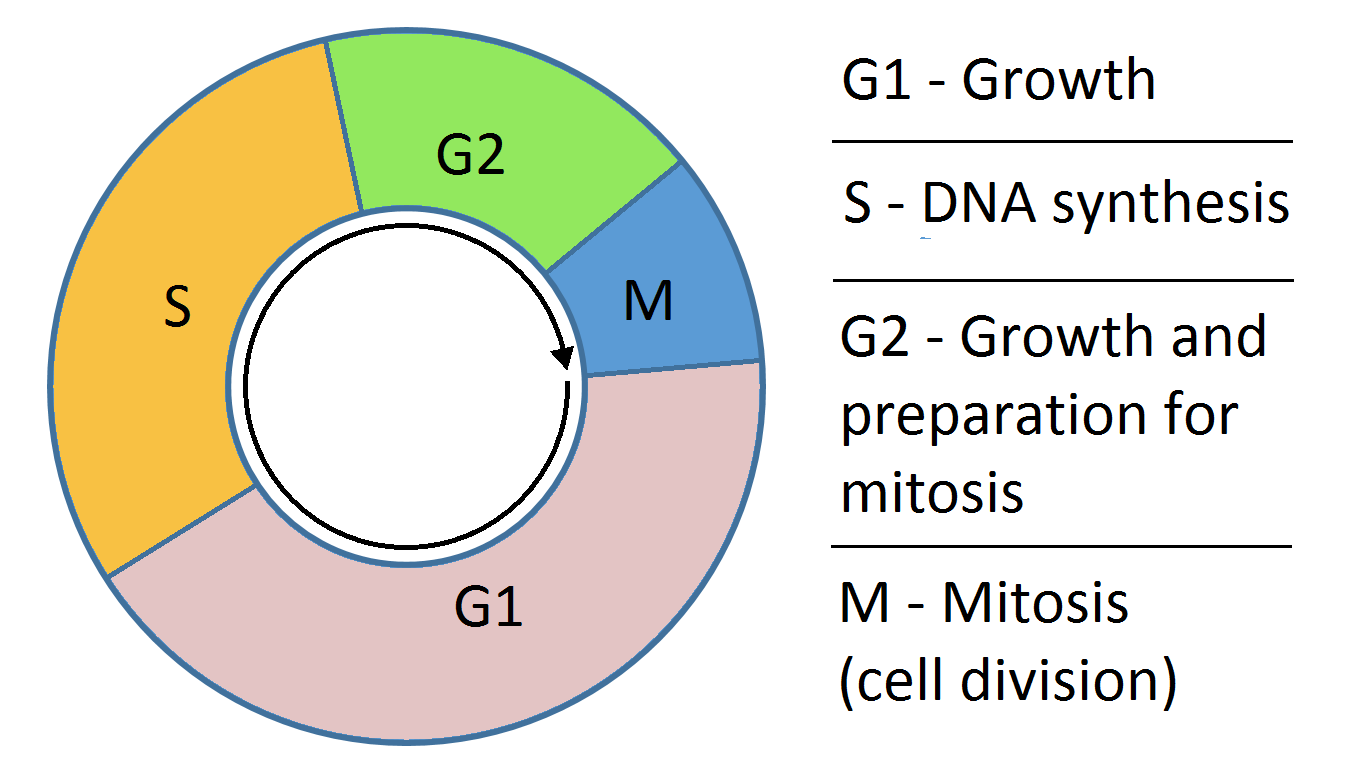
Figure 3.5.1
Figure 3.5.1 is a simplified representation of the typical cell cycle. Note that there are four consecutive phases starting with G1. The colors indicate the approximate portion of the cycle each phase requires.
The cell cycle is divided into four phases as illustrate in Figure 3.5.1. All together, the first three phases, Growth 1, Synthesis, and Growth 2 , represent Interphase. Interphase takes up about 90% of the cell cycle in terms of time. The Growth 1 (G1) phase directly follows cytokinesis, the physical separation of cells after mitosis has taken place. During this phase, the cell is creating messenger RNA and proteins. The Synthesis (S) phase begins with the replication of the DNA within the cell. This requires the presence of enzymes and nucleotides and results in the cell having twice as much DNA as it had started with. The cycle continues into Growth 2 (G2) phase where proteins and other materials continue to be created in preparation for mitosis. This phase ends as mitosis begins.
ACTIVITY:
- Watch this video clip (about 30 seconds) and write down your observations. Feel free to pause or review it to complete your observations.
- Answer the following questions based on your observations.
- How many cells did you observe go through mitosis?
- How many cells were present at the end of mitosis?
- The red/orange structures in the video of the cell are DNA. Briefly describe any changes you noticed with the DNA through the process of mitosis.
- Describe what you think the function is of the neon green structure in the cell.
- This video is about 30 seconds in length. Do you think this represent the actual time this process requires? Explain your reasoning.
- Share your work as directed by your instructor.
Remember that mitosis is part of the larger cell cycle. It is not a process that just happens. In order to be able to go through mitosis, the cell must go through G1, S, and G2. The timing of these processes leading up to mitosis is tightly controlled by proteins within the cell. The signal protein for mitosis is produced in the G2 phase and triggers M phase.
[1] Cell_cycle_simple by Simon Caulton / CC BY-SA 3.0